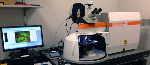
What IS that One-of-a-Kind Microscope?
What IS That? A custom Renishaw Raman Microscope with dual laser system coupled to a Leica Upright Microscope and LED fluorescence excitation and emission source system. We believe that this is the only microscope in the world with this custom configuration.

What does it do? It processes data and analyzes images in “real-time” and allows us to create 2-D image and 3-D images of cancer cells and tumors in their native environment in a non-invasive manner using the spectral data.
How will we use it? Professor Michael Fenn and Professor Larry Hench of the Biomedical Engineering department will be using it for novel research in areas such as new cancer diagnostics and treatments, improved understanding of tumor and cancer cell biology for anti-cancer agent development, monitoring cellular interactions with a new generation of bioactive materials for bone regeneration, discovery of new early-stage biomarkers for breast cancer, cardiovascular pathologies and tissue engineering construct development.

What do we hope to do with our findings? One of the main objectives will be to develop ways to both simultaneously diagnose and treat cancers in real-time. This is called “theranostics” as it is diagnostics and therapy combined into one platform. This will open up the ability to do microscopic examination of living tissue inside the body during surgical oncology applications, as well as improved histopathology capabilities, diagnostic sensitivity, and the greatly improved understating of complex biological mechanisms.

Where can I find it on campus? On the third floor of the Link building in the Biomedical Engineering department.
If you’re interested in projects like these, you should consider an undergraduate or graduate degree in biomedical engineering.